- On this page: Does My Doctor See?
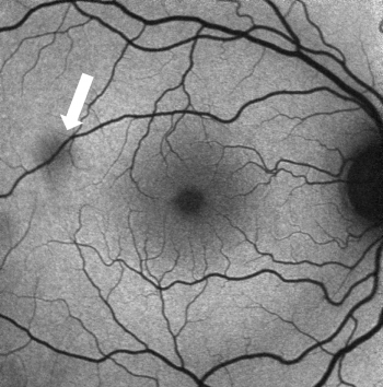
Fundus autofluorescence (FAF) is a non-invasive diagnostic test that involves taking digital photographs of the back of the eye without a contrast dye.

Autofluorescence imaging of the retina can help with the early detection of retinal disease in serious eye conditions such as:
Fluorophores are chemical structures that possess fluorescent properties when exposed to light of a particular wavelength. Lipofuscin is a fluorophore that accumulates with age but can also occur from cell dysfunction. Lipofuscin autofluorescence, then, refers to the natural ability of lipofuscin to glow when excited with short to medium wavelengths of visible light.
Fundus autofluorescence imaging records fluorescence that occurs naturally in the eye or accumulates as a result of disease. FAF uses light to produce images rather than radiation, sound, or radiofrequency waves. As a result, there is no risk to the patient or harmful side effects.
Your doctor can evaluate the health of the retina using this FAF imaging technology. This technology allows your doctor to detect diseases of the retina at early stages and has made timely intervention possible for many retinal conditions.
Fundus autofluorescence imaging may be performed multiple times to monitor the progression of retinal diseases, the need for intervention, and the response to treatment.