- On this page: Symptoms
- Flashes and Floaters
- Causes and Associations
- Testing
- Treatment
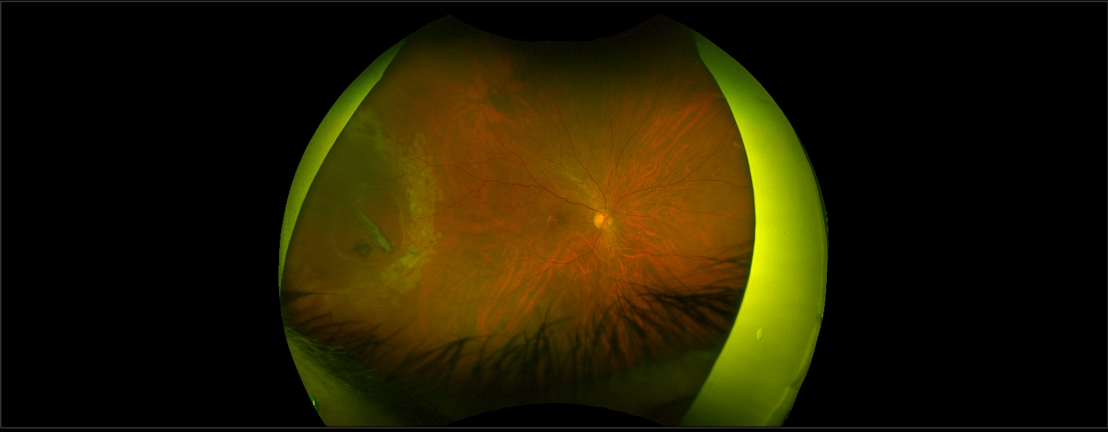
A retinal tear occurs when the retina begins to tear away from its normal position. Retinal tears can lead to a retinal detachment which requires immediate treatment to prevent vision loss. There is no way to predict a retinal tear.
Retinal tears are often a result of Posterior Vitreous Detachment (PVD), which is a natural change that occurs as the eye ages. With PVD, the vitreous gel that fills the eye becomes liquid and condenses, eventually separating from the retina. If the vitreous pulls too hard on the retina during this process, it can cause a retinal tear.

Symptoms of a retinal tear include:
If there is bleeding into the vitreous gel (vitreous hemorrhage), vision may become very blurry as the blood disperses throughout the gel.
Retinal tears can progress to a retinal detachment as fluid enters through the tear and underneath the retina. This causes the retina to peel off the back wall of the eye, resulting in a shadow that typically starts in the peripheral vision. This shadow can enlarge as the retina continues to detach and eventually leads to blurry and/or distorted central vision.
Flashes
Light flashes are a result of retina stimulation and often occur when the vitreous gel pulls on or separates from the retina. This phenomenon can also originate from the visual centers of the brain, like visual auras that are associated with migraine headaches.
Floaters
Floaters are small clumps of cells or tissue that form in the vitreous gel, the clear jelly-like substance that fills the inside cavity of the eye. They may appear as little dots, circles, lines, or cobwebs. Uncommonly, floaters are the result of inflammation within the eye or from crystal-like deposits that form in the vitreous gel.
Are Flashes and Floaters Serious?
Flashes and floaters are symptoms of PVD. During or after PVD, the retina or a blood vessel may be torn, resulting in a retinal tear or bleeding in the vitreous. An increase in the frequency or severity of flashes and floaters is concerning for the development of a retinal tear or bleed in the vitreous.
Without an examination by an ophthalmologist, symptoms alone cannot determine whether floaters or flashes are caused by PVD or a retinal tear. Any sudden onset of new floaters or flashes should be evaluated promptly.
The vitreous gel deteriorates with age and it can separate from the retina, resulting in a tear. Retinal tears are more common in those 50 years and older.
Other potential risk factors of retinal detachment include:
A comprehensive eye examination is important to assess retinal tears, including vision testing, eye drops to dilate the pupils and a complete examination of the front and back of the eye. It is best if a driver accompanies the patient during the eye exam as pupillary dilation may create near-vision blurring.
If a retinal tear is detected prior to the development of a large retinal detachment, laser or retinal cryopexy is often successful in preventing a detachment.
Laser Photocoagulation
Laser photocoagulation is performed in an office setting. A laser delivers split-second bursts of intense light energy to create a seal around the retinal tear or small retinal detachment. If there is too much bleeding in the vitreous gel, laser photocoagulation cannot be performed.
Retinal Cryopexy
Retinal cryopexy is a procedure performed in an office setting that utilizes intense cold to freeze portions of the retina. The freezing is performed around the retinal tear and promotes the formation of a seal around a retinal tear.
If you have any signs or symptoms of retinal tears, contact us today to meet with a Mid Atlantic Retina specialist. Our team can work with you to protect your vision and minimize your complications.
Being diagnosed with a retinal tear can be frightening. While it is a serious condition...